

When Superficial Fascia Goes Deep
Superficial fascia may not get the same attention as deep fascia, but it plays an extremely important role in the organization of tissues and the structure of the body.
The human body uses various systems to absorb impact and cope with uneven surfaces while maintaining a stable base of support. Anatomical structures in the foot, knee, pelvis, and spine all work in tandem to distribute forces and prevent wear and tear on the body as it interfaces with other objects—such as the ground—during gait. Much like the suspension of a car, an assembly of rigid links and optimally mobile joints function as a kinematic chain to manage forces and movement in, and through, weight-bearing structures.
Focusing on the pelvic girdle, parallels can be made between the springs, shock absorbers, and linkages of a car's suspension and the bones, joints, and ligaments functioning as a kinematic chain in this region. The anterior pelvic girdle is formed by the pubic symphysis, a cartilaginous joint uniting where the two pubic rami join at the midline. Posteriorly, the pelvic girdle consists of two large, stable synovial joints where the large right and left ilia join the centrally suspended sacrum. The weight of the torso and upper body descends through the spine, pelvic girdle, and then laterally through the femoral neck on each side, and finally through the lower extremities.
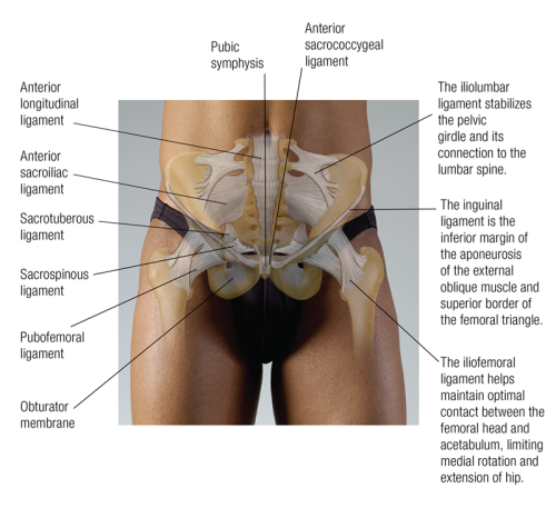
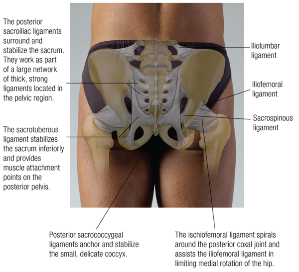
Distribution of weight in the pelvic girdle primarily occurs in the posterior region. The sacrum is suspended between the two large ilia by the posterior sacroiliac ligaments, and symmetrical tension in these ligaments is key to maintaining alignment and stability in the pelvic girdle. A complex network of ligaments, including the sacroiliac, sacrotuberous, and sacrospinal ligaments, maintains the position of the sacrum, much like springs maintain the position and equal tension on the fabric at the center of a trampoline. Balanced tension in this ligamentous network maintains optimal deflection (the degree to which a structural element is displaced under a load) and spring rate (the change in force exerted divided by the change in deflection) in the pelvic girdle. The resulting mechanical alteration of forces helps decrease impact on the soft tissues and articulating surfaces further up the kinematic chain, particularly in the spine.
Slight mobility provided anteriorly by the cartilaginous disc at the pubic symphysis, in conjunction with the two sacroiliac joints located posteriorly, accommodates forces generated with the asymmetrical weight transfer and rotational motion of the sacrum during gait. It also assists in maintaining a relatively constant torso and head position, allowing the eyes to remain level with the horizon, by minimizing body lean as weight shifts from one foot to the other. The combination of posterior stability and slight anterior mobility makes the pelvic girdle an efficient and effective suspension system capable of both intense, and subtle, force distribution at the center of the body.
Proper function of the pelvic girdle suspension system relies on a balance between rigidity and mobility. Excessive joint mobility due to ligament injury or general hypermobility may lead to chronic misalignment and excessive wear on joint surfaces. Reduced mobility decreases the efficacy of the suspension system, transferring potentially harmful forces further up the kinematic chain. Trauma and chronic degeneration to the intervertebral discs and other essential structures may result as they are forced to cope with excessive compression. Asymmetrical force distribution due to leg-length discrepancy, functional scoliosis, or other chronic use patterns can prove equally challenging. Encouraging optimal postural alignment and functional mobility, maintaining symmetric stability both statically (ligamentous) and dynamically (muscle tension patterns), and effective activation of core muscles all help support the function of the pelvic girdle suspension system.
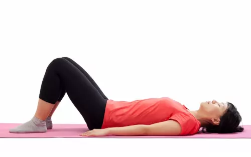
Lie on your back with your knees bent and feet flat on the floor.
Press your low back flat into the floor by tightening your abdominal muscles.
Make sure your glutes and hamstrings stay relaxed. Breathe normally.
Relax your abdominals and let the curve in your back return.
Practice flattening and restoring the curve by tightening and relaxing your abdominal muscles.
Editor's note: The Client Homework element in Functional Anatomy is intended as a take-home resource for clients experiencing issues with the profiled muscle. The stretches identified in Functional Anatomy should not be performed within massage sessions or progressed by massage therapists, in order to comply with state laws and maintain scope of practice.
Superficial fascia may not get the same attention as deep fascia, but it plays an extremely important role in the organization of tissues and the structure of the body.
Understanding tendons—their shapes, lengths, and organization—improves an MT’s touch vocabulary and facilitates a more skilled touch.
While the neck is a bridge, a pathway, the position of the neck and head can also indicate a multitude of other things happening beneath the surface.
Understanding fibroblasts and the extracellular matrix changes how we think about the tissue we touch.